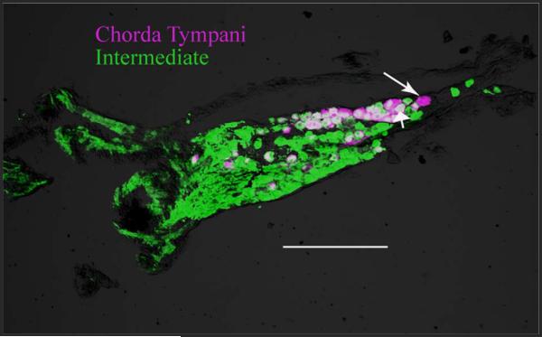
Figure 9.
Photomicrograph of a 20-μm section through a geniculate ganglion showing the cells that were labeled at the time of the CTX (DiI-labeled, magenta cells) and all geniculate ganglion cells that send a process centrally through the intermediate nerve (BDA reacted with streptavidin 488, green cells) at 30 days post-CTX. Most (~95%) DiI-labeled cells were also labeled with BDA (double labeled, white), indicating that the chorda tympani neurons that had a peripheral axon at the time of CTX maintained their central process. The long arrow points to a magenta-only cell, and the short arrow points to a double-labeled cell (white cell). Scale bar = 200 μm.

