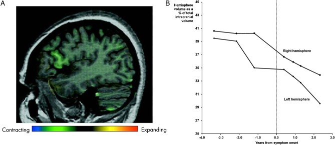
Figure 1.
MRI changes in the proband: (A) sagittal MRI showing focal anterolateral left frontal lobe atrophy, particularly centred around the pars opercularis, using voxel compression mapping between the first and second scans (3.4 and 2.1 years prior to symptom onset) (reprinted from Janssen et al,1; (B) changes in left and right hemispheric volume over time.

