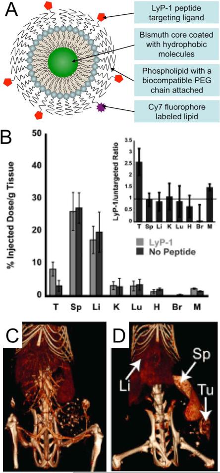
Figure 7.
A) Schematic depiction of the structure of a micelle-based, targeted bismuth core nanoparticle. B) Biodistribution of targeted and non-targeted nanoparticle formulations at 24 hours post-injection. T=tumor, Sp=spleen, Li=liver, K=kidney, Lu=lungs, H = heart, Br = brain, M = muscle. CT images acquired C) immediately and D) 24 hours post-injection of targeted nanoparticles. Figure adapted with permission from (48).

