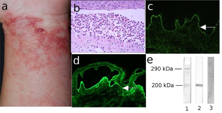
Fig. (12).
Anti-p200 pemphigoid (a) Erythema, blisters, erosions and crusts in a 53-year old patient with anti-p200 pemphigoid. (b) Histopathological examination reveals subepidermal cleavage and a neutrophil-rich inflammatory infiltrate. (c) Direct immunofluorescence (IF) microscopy analysis of perilesional skin shows linear IgG deposition at the dermo-epidermal junction. (d) Serum IgG autoantibodies bind to the dermal side of 1M NaCl-split skin by indirect IF microscopy (all magnification 200x). (e) Dermal extracts were separated by 6% SDS-PAGE, transferred on nitrocellulose and immunoblotted with serum from patients with epidermolysis bullosa acquisita (EBA; lane 1), anti-p200 pemphigoid (p200; lane 2) and normal human serum (NHS, lane 3).

