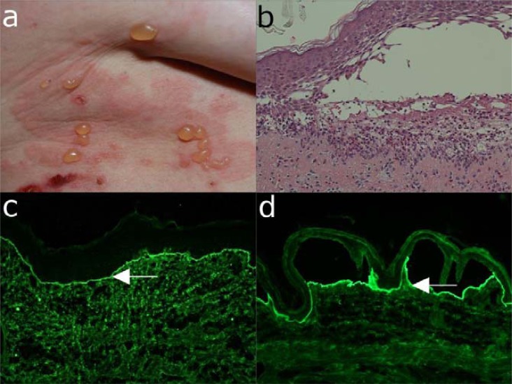
Fig. (13).
Diagnostic features of epidermolysis bullosa acquisita (EBA). (a) Clinical picture of a 61-year old female patient with the inflammatory form of EBA showing erythema, tense blisters, erosions and crusts on the lateral abdomen. (b) Histopathology analysis of the lesional skin shows dermal-epidermal separation and a neutrophil-rich inflammatory infiltrate. (c) Direct immunofluorescence microscopy of perilesional skin reveals deposits of IgG along the basement membrane zone. (d) Indirect immunofluorescence microscopy on 1M NaCl split-skin shows binding of IgG autoantibodies to the dermal side of the dermal-epidermal junction (magnification 200x).

