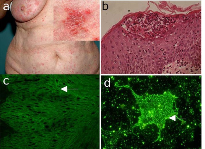
Fig. (6).
IgA pemphigus. (a) Clinical picture of a 56-year old woman with IgA pemphigus showing pustules on the abdomen. Inset: close-up view showing pustules, blisters, erosions, and crusts on an erythematous background. (b) Histopathological examination reveals subcorneal acantholysis with an inflammatory infiltrate consisting mainly of neutrophils. (c) IgA autoantibody binding with an intercellular pattern on monkey esophagus by indirect immunofluorescence (IF) microscopy. (d) Indirect IF microscopy using COS-7 cells transfected with desmocollin 1-cDNA as substrate reveals autoantigen-specific IgA serum autoantibodies.

