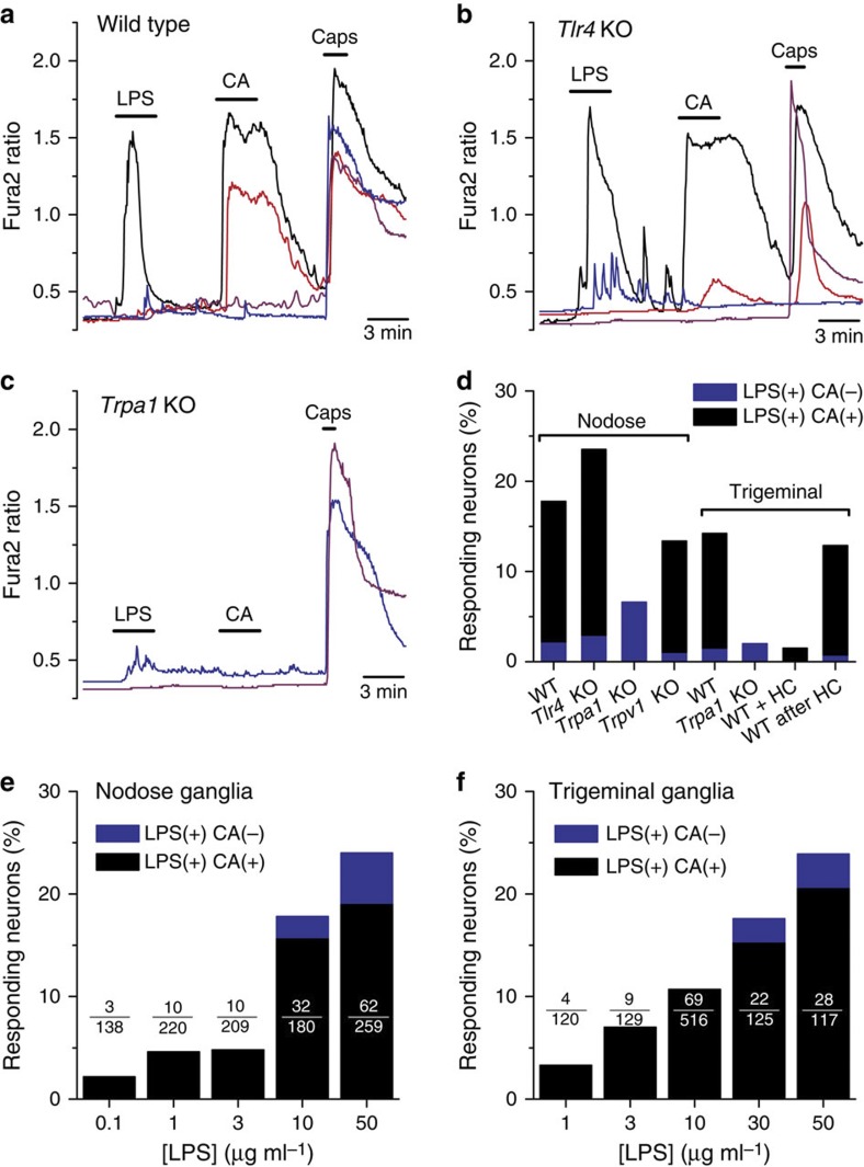
Figure 1. TRPA1 mediates stimulation of nociceptor neurons by LPS.
(a–c) Representative examples of the effects of LPS (10 μg ml−1) on [Ca2+]i levels in nodose ganglion neurons isolated from WT (a), Tlr4 KO (b) and Trpa1 KO mice (c). Cinnamaldehyde (CA, 100 μM) and capsaicin (Caps, 100 nM) were applied to identify TRPA1- and TRPV1-expressing neurons, respectively. (d) Percentage of mouse nodose and TG sensory neurons responsive to LPS (blue) or to LPS and CA (black). The labels ‘WT+HC’ and ‘WT after HC’ refer to the responses to LPS (10 μg ml−1) observed in the presence of the TRPA1 inhibitor HC-030031 and after its removal, respectively. (e,f) Percentage of nodose (e) or TG (f) neurons responding to LPS (blue) or to LPS and CA (black) as a function of LPS concentration.

