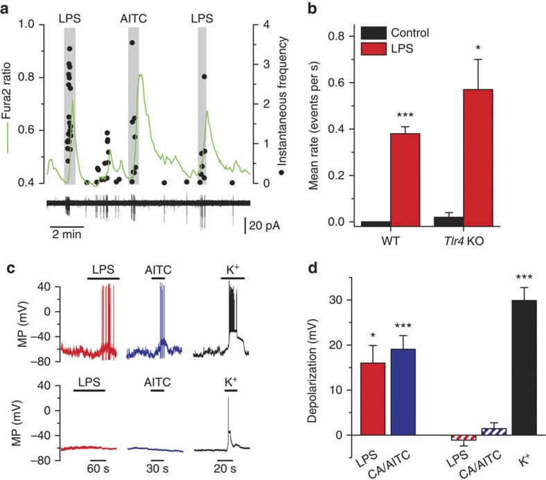
Figure 2. E. coli LPS excites TRPA1-expressing sensory neurons.
(a) Simultaneous recording of [Ca2+]i level (green trace) and instantaneous firing frequency (black dots) in cell-attached mode in a nodose neuron from a Tlr4 KO mouse. The large calcium transients (upper graph) during LPS and MO application correlate with the firing of action potentials (lower graph). (b) Summary results of effects of E. coli LPS (10 μg ml−1) on mean±s.e.m. firing rate in nodose neurons from WT (n=4) and Tlr4 KO mice (n=3), (paired t-test). WT neurons were electrically silent before LPS application. (c) Examples of recordings of membrane potential in the perforated-patch current-clamp mode (Ihold=0 pA) in two nodose neurons. LPS (10 μg ml−1) induces rapid depolarization and firing of action potentials in the TRPA1-expressing neuron (that is, AITC positive; upper traces). In contrast, LPS had no effect on membrane potential in the TRPA1-negative neuron (lower traces). (d) Average±s.e.m. depolarization of the resting membrane potential produced by LPS (n=5) and AITC (AITC) or CA (n=10) in TRPA1-expressing neurons (solid bars). The dashed bars represent the average depolarization produced by the application of LPS, AITC/CA or high extracellular potassium (60 mM) in neurons not expressing TRPA1 (n=10). Statistically significant differences of membrane potential (relative to control) are denoted by *P<0.05 and ***P<0.001, paired t-test).

