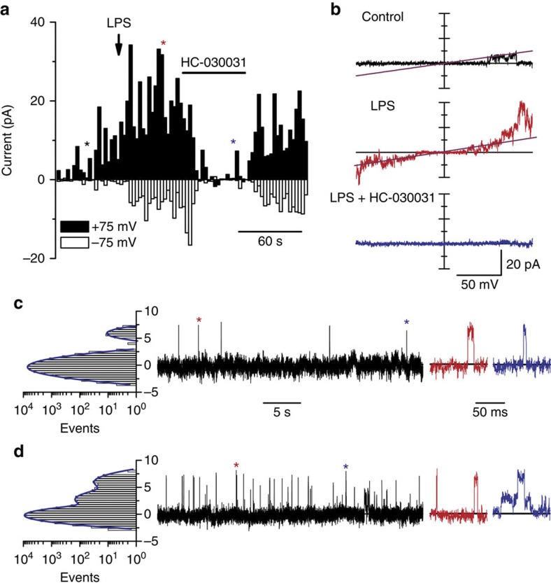
Figure 4. LPS activates TRPA1 channels in a membrane-delimited manner.
(a) Time course of current amplitudes recorded in outside-out patches of TRPA1-expressing CHO cells at –75 and +75 mV. Currents were elicited by voltage ramps from −100 to +100 mV from a holding potential of 0 mV. The arrow indicates the moment of application of LPS (10 μg ml−1) and the horizontal bar indicates the period of exposure to 50 μM HC-030031. The coloured asterisks mark the time points at which the corresponding current traces shown in b were recorded. Single-channel records in the outside-out configuration at +75 mV, in control solution (c) and in the presence of 10 μg ml−1 LPS (d). Events marked with an asterisk are displayed at an expanded timescale. The histograms on the left show the corresponding distributions of the current amplitudes. All records in the figure were obtained in Ca2+-free conditions.

