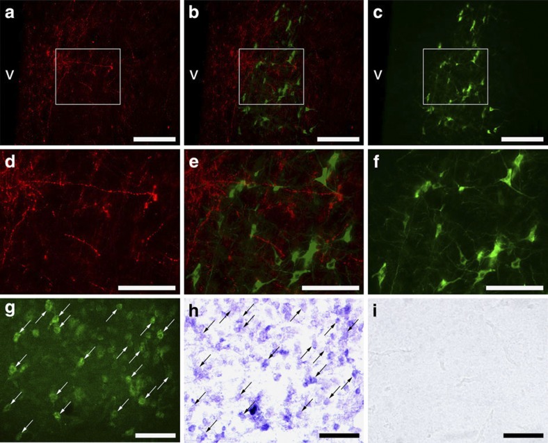
Figure 3. Distribution of GnIH-ir fibres and aromatase-ir cells and GnIH receptor mRNA in the POA.
(a) Abundant GnIH-ir neuronal fibres were observed in the POA. v, third ventricle. Bar, 100 μm. (c) A cluster of aromatase-ir cells was observed in the POA. v, third ventricle. Bar, 100 μm. (b) The merged image of the pictures (a) and (c) showed abundant GnIH-ir neuronal fibres in the vicinity of aromatase-ir cells. v, third ventricle. Bar, 100 μm. Similar results were obtained in repeated experiments using four different birds. (d–f) Higher magnification of the blocked area in (a–c). Bar, 50 μm. (g–i) Aromatase immunohistochemistry (g) and in situ hybridization for GnIH receptor (GPR147) mRNA on the same sections showed that almost all aromatase-ir cells express GPR147 mRNA. Arrows in (g) and (h) indicate identical cells in the POA. Bar, 50 μm. In situ hybridization using sense RNA probe (i) served as controls. Similar results were obtained in repeated experiments using three different birds.

