Figure 4.
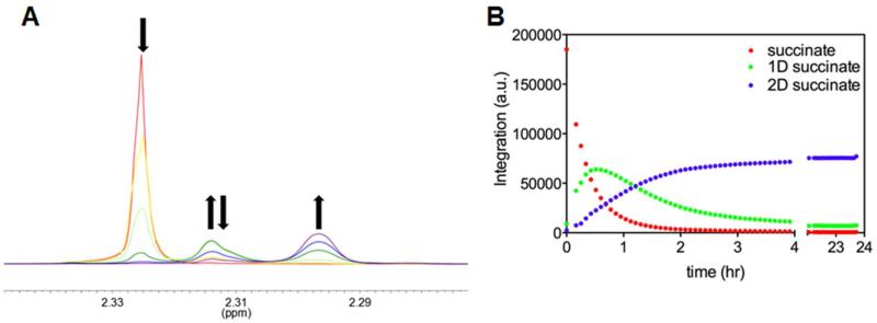
“Time Course of Succinate 1H Resonances.” A) The red scan was taken before 1 μM ICL was added, the yellow 10 min after ICL addition, light green after 104 min, dark green after 329 min, blue after 659 min, and purple after 1313 min. B) Integration of each isotopic form of succinate after the addition of 5 μM ICL over 23.7 hours. Time zero is before ICL addition.

