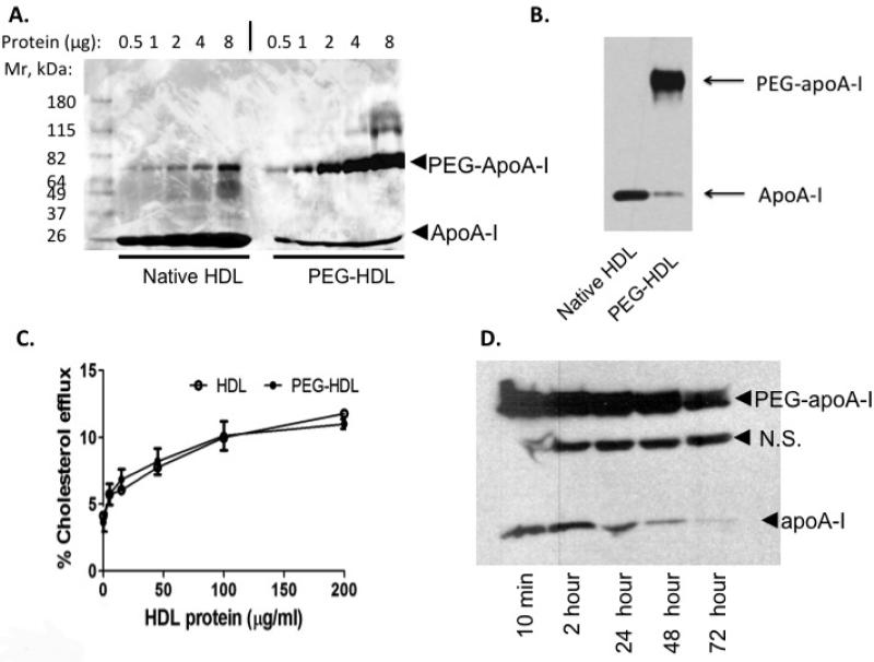
Figure 2. Characterization of pegylated human HDL.
(A) HDL with or without pegylation using M-PEG-ALD 20K at 4°C for 24 hours and analyzed by SDS-PAGE and Coomassie Blue staining. The minor higher Mr band in native HDL samples is likely albumin in the HDL preparations. (B) Western analysis using an anti-apoA-I antibody to HDL and PEG-HDL. (C) Cholesterol efflux for 3 hours from cholesterol-loaded macrophage cells derived from THP-1 cells to HDL or PEG-HDL prepared as in (A). (D) Plasma clearance of PEG-HDL following injection of 40mg/kg PEG-HDL prepared as in (A) and analyzed by Western analysis. N.S.= non-specific band.

