
Figure 3. Characterization of pegylated human rHDL.
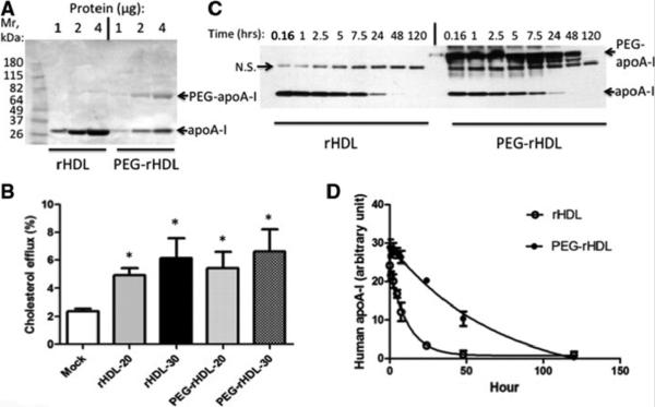
(A) rHDL pegylated with M-PEG-ALD 20K at 4°C for 16 hours and analyzed by SDS-PAGE and Coomassie Blue staining. (B) Cholesterol efflux for 3 hours from cholesterol-loaded LXR activated THP-1 macrophages to 20 or 30 μg rHDL or PEG-rHDL protein/ml. (C) Plasma clearance of rHDL or PEG-rHDL following injection of 60mg/kg rHDL or PEG-rHDL protein into circulation and analyzed by Western analysis. N.S.= non-specific band. (D) Densitometry quantification and analysis of (C), n=4.