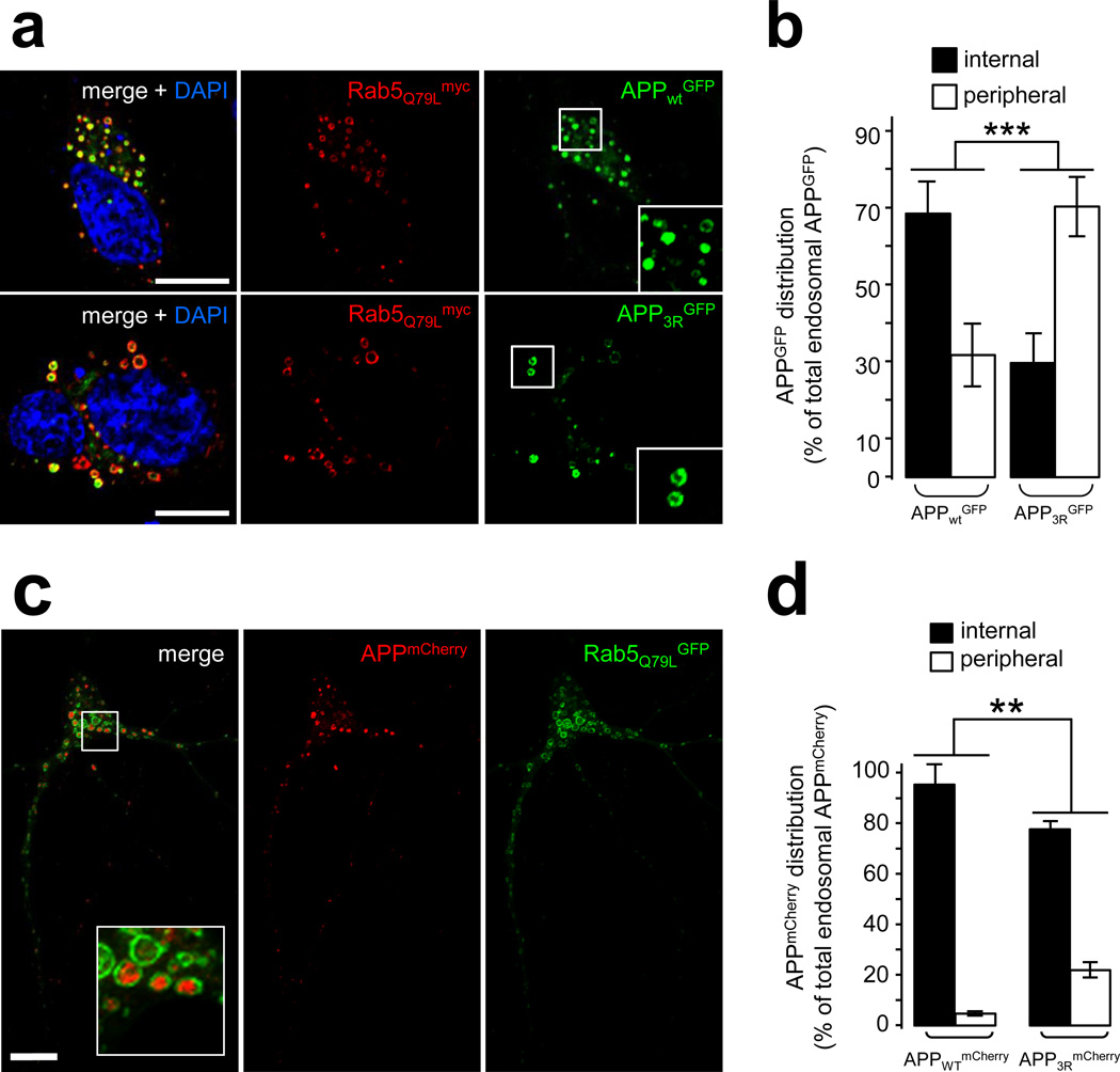
Figure 6. Blocking the intraluminal sorting of APP enhances its amyloidogenic processing.
a) Confocal analysis of HeLa cells transfected with Rab5Q79Lmyc and APPWTGFP (top panel) or APP3RGFP (middle panel). Cells were labeled with an anti-myc antibody (red) and counterstained with DAPI (blue). Scale bar = 10µm.
b) Bar diagram showing the quantification of the endosomal distribution of human APPGFP expressed as % of total endosomal APPGFP: internal = inside endosomal lumen, peripheral = limiting membrane of the endosomes. Values denote means ± SEM (for each construct, n = 18 cells from 3 experiments with an average quantification of 15 endosomes per cell). ***, denotes P values < 0.001 from a Student's t-test.
c) Confocal analysis of mouse hippocampal neurons transfected at DIV9 with Rab5Q79LGFP and APPWTmCherry and fixed after a 24 h treatment with γ-secretase inhibitor XXI. Scale bar =10 µm. The inset shows APP-containing endosomes.
d) Bar diagram showing the quantification of the endosomal distribution of human APPWTmCherry and APP3RmCherry in hippocampal neurons processed as described in (c). The APPmCherry distribution is expressed as % of total endosomal APPmCherry: internal = inside endosomal lumen, peripheral = limiting membrane of the endosomes. Values denote means ± SEM (n=45 and 18 cells for APPWTmCherry and APP3RmCherry, respectively from 3 experiments with an average quantification of 20 endosomes per cell). **, denotes P values < 0.01 from a Student's t-test.

