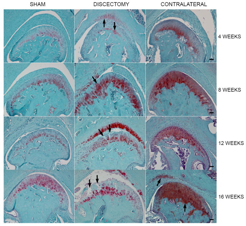
Figure 1.

Histology of TMJs in mice. Each image shown in the figure is a representative section for each experimental group. Morphological changes of articular cartilage degeneration were seen in the discectomy mice, such as increased proteoglycan staining at early stage, chondrocyte clusters, reduced proteoglycan staining at late stage, fibrillation and the loss of articular cartilage. The morphological change was also seen in the contralateral non-surgical TMJs. However, only increased proteoglycan staining was present in the articular cartilage of the condyle at early stage and the increased staining was extended to the fossa of temporal bone at late stage. The morphological changes in each section were observed in all joints in each animal group. (Bar=50 μm) Table 1 Average scores for the sham, discectomy and contralateral non-surgical TMJs.
As the mice continued to age, the scores of the discectomy and contralateral non-surgical joints continued to increase, however, the rate of score increase in the non-surgical groups was significantly slower, compared to that in the discectomy groups
