
Figure 1.
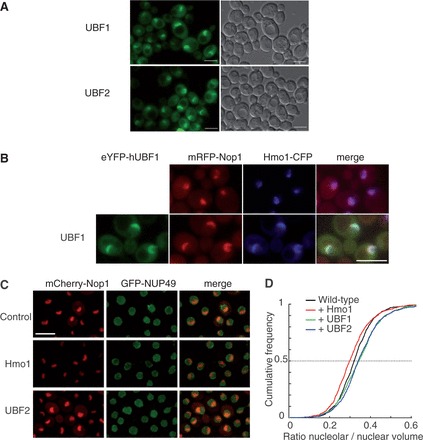
When expressed in S. cerevisae, UBF1 and UBF2 are nucleolar and increase nucleolar volume. (A) UBF1 and UBF2 fused to YFP and produced in yeast concentrate in a nuclear crescent shape structure. (B) The construct encoding the YFP-UBF1 fusion protein (green) was used to transform a strain producing a Hmo1-CFP fusion protein (blue) and an mRFP-Nop1 fusion protein (red), which define different subdomains of the nucleolus. UBF1, Hmo1and Nop1 fully co-localize. (C) A strain producing mRFP-Nop1 fusion protein (red) and GFP-Nup49, which reveals the nucleolus and nuclear, respectively, periphery was transformed with empty plasmid (Control), a plasmid over-expressing Hmo1 or UBF1 (not shown) or UBF2. (D) Nuclear and nucleolar volumes were imaged by confocal microscopy and quantified using automated detection software (32). Cumulative frequency plots of the nucleolar to nuclear volumes ratio show a decrease on Hmo1 overexpression [Two-sample Kolmogorov–Smirnov test (KS), P = 2.7e−06] and a significant increase on UBF1 (KS test, P = 1.6e−08) or UBF2 (KS test, P = 4.3e−07) expression relative to control. Scale bar is 5 µm.