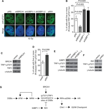
Figure 7.

The formation of IR-induced (pT371)TRF1 foci requires BRCA1 but is inhibited by 53BP1 and Rif1. (A) Indirect IF with anti-pT371 antibody on 12 Gy IR-treated HeLaII cells depleted for BRCA1 or 53BP1. Knockdown of 53BP1 was performed with two independent shRNA (sh53BP1-1 and sh53BP1-2). Cell nuclei were stained with DAPI in blue. (B) Quantification of the percentage of cells with five or more IR-induced (pT371)TRF1 foci in HeLaII cells depleted for BRCA1 or 53BP1 from (A). Standard deviations from three independent experiments are indicated. (C) Western analysis of HeLaII cell extracts with or without depletion of BRCA1. Immunoblotting was performed with anti-BRCA1, anti-pT371, anti-TRF1 or anti-γ-tubulin antibody. (D) Quantification of the percentage of cells with five or more IR-induced (pT371)TRF1 foci in HeLaII cells knocked down for Rif1. Standard deviations from three independent experiments are indicated. (E) Western analysis of HeLaII cell extracts with or without depletion of 53BP1. Immunoblotting was performed with anti-53BP1, anti-pT371, anti-TRF1 or anti-γ-tubulin antibody. (F) Western analysis of HeLaII cell extracts with or without depletion of Rif1. Immunoblotting was performed with anti-Rif1, anti-pT371, anti-TRF1 or anti-γ-tubulin antibody. (G) Model for the role of (pT371)TRF1 in facilitating HR and checkpoint activation. See the text for more information.
