
Figure 4.
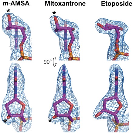
The unbiased difference electron density maps (mFo-DFc, contoured at 4.5σ) of the −1 nucleotides are shown to validate the ring puckering change in the deoxyribose in the presence of the respective drugs. The m-AMSA- and mitoxantrone-stabilized axial positions of 3′-OH are indicated by black stars.