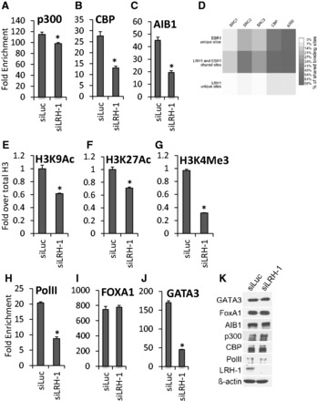
Figure 5.

LRH-1 is required for co-activator loading and histone modification. MCF-7 cells were transfected with LRH-1 siRNA. ChIP was performed using antibodies for ERα cofactors (A–C, I and J), histone H3 acetylation and methylation marks (E–G) or PolII (H), followed by real-time PCR of the pS2 ERE, as shown. Enrichment is shown relative to the IgG control (n = 3). The acetylated and methylated H3 ChIP was first normalized to total H3, then to the IgG control. Errors bars = SEM, *P < 0.05. (D) Heatmap of ERα co-activator binding [data from (71)], showing the percentage of overlapping SRC1, SRC2, SRC3, CBP and p300 binding events at LRH-1 and ERα unique and shared sites. (K) Western blotting of the lysates in parts (A–C, E–J), is shown.
