
Figure 1.
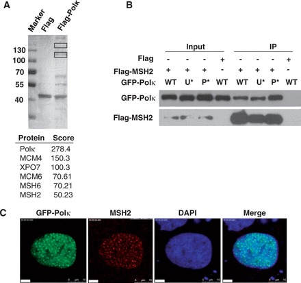
MSH2 physically interacts with Polκ. (A) Identification of MSH2 as a Polκ-associated protein. HEK 293T cells were transfected with a 3× Flag-Polκ expression vector and UV irradiated (15 J/m2). The cells were cross-linked, and lysates were harvested at 4 h after UV irradiation and immunoprecipitated with an anti-Flag M2 agarose affinity gel as described in ‘Materials and Methods’ section. The affinity-purified proteins associated with FLAG-Polκ were separated by SDS–PAGE and revealed by silver staining. The bands (indicated with black rectangles) were excised and analyzed via mass spectrometric sequencing. The results from mass spectrometry analysis were shown below. (B) Co-IP experiment showing the interaction between MSH2 and Polκ. Flag or Flag-MSH2 and GFP-Polκ or its mutants were co-transfected into 293T cells, and the lysates were immunoprecipitated using an anti-Flag M2 agarose affinity gel. The immunoprecipitate was then examined via western blot using anti-GFP antibody (Top panel) or anti-Flag antibody (Bottom panel). The input included 2% of the cell lysate used. WT represents wild-type GFP-Polκ, U* represents GFP-Polκ-UBZ*, P* represents GFP-Polκ-PIP*. (C) Co-localization of MSH2 and GFP-Polκ after UV irradiation. GFP-Polκ-transfected U2OS cells were UVC irradiated (15 J/m2) and further incubated for 8 h. The cells were then fixed and immunostained with anti-MSH2 monoclonal antibody and DAPI-stained.