Figure 3.

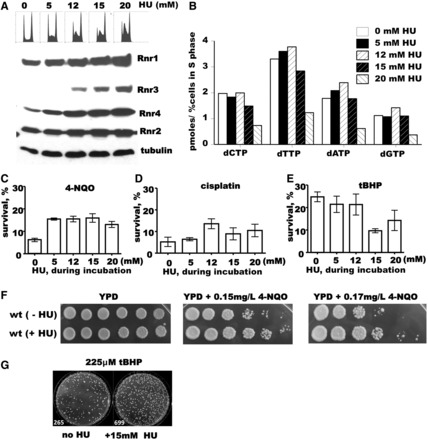
Low concentration of HU improves DNA damage tolerance of the wild-type strain. Western blot analysis of Rnr1–4 protein levels and corresponding flow cytometry histograms (A) and dNTP concentration measurement (B) in the wild-type strain (W1588-4C) after 10 h incubation with 0, 5, 12, 15 or 20 mM HU. (C–E) Analysis of DNA damage tolerance of the wild-type strain incubated 10 h with 0, 5, 12, 15 or 20 mM HU. Cells were spun, washed once with water, and appropriate dilutions were plated on YPD or YPD containing 0.15 mg/l 4-NQO (C), 2 mMcisplatin (D) or 325 μM t-BHP (E). (F) Spot assays on YPD media containing 0.15 mg/l or 0.17 mg/l 4-NQO were performed using 1:10 serial dilutions of wild-type cells pre-incubated 10 h with 0 mM HU (top row) or 12 mM HU (bottom row). (G) Analysis of DNA damage tolerance. Wild-type cells (W1588-4C) were grown to mid-log phase and appropriate dilutions were plated on YPD containing 325 μM t-BHP with or without 15 mM HU. The number of colonies is shown.
