
Figure 3.
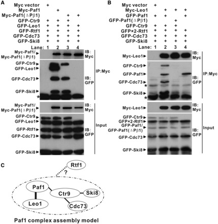
Assembly of human PAF1C. (A and B) Co-IP experiments of PAF1C formation by Paf1 (A) and Leo1 (B). Extracts were prepared from HEK293T cells transfected with various combinations of plasmids as indicated, immunoprecipitated with agarose-conjugated anti-Myc and subsequently immunoblotted with anti-Myc (upper top panels) or anti-GFP (lower bottom panels) as indicated. The top panel shows the IP results. The bottom panel shows 20% of the GFP fusion proteins for each IP. Nonspecific bands and the heavy chain of the antibody are marked by a star and asterisk, respectively. (C) Model of PAF1C assembly. The bold line represents the interaction in the crystal structure of the Paf1(161–250)-Leo1(370–462) heterodimer. The fine lines represent the interaction obtained from the IP results. The dotted line represents interactions that we could not detect in the IP experiments.