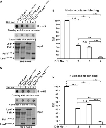
Figure 5.

Interaction of the Paf1/Leo1 subcomplex with histone octamer and nucleosome. (A and C) Dot-blot overlay assays of the Paf1/Leo1 complex and histone octamer and nucleosome. The bound histone octamer or nucleosome was detected by immunoblotting with antibody against histone H3 (top panel). The PVDF membrane was stained with Coomassie blue (middle panel). Each recombinant Paf1/Leo1 complex was resolved by SDS-PAGE to normalize the sample inputs (bottom panel). (B and D) Bar graph of the binding histone octamer and nucleosome. The error bars indicate the standard error of the mean (n = 3, separate experiments). n.s., not significant (P > 0.05). **P < 0.01, ***P < 0.001.
