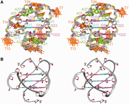
Figure 4.

Stereoviews of the htel27[Br22] G-quadruplex structure in Na+ solution. (A) Ten superimposed refined structures. (B) Ribbon view of a representative structure. Anti guanines are colored in cyan; syn guanines and 8-bromoguanine, magenta; adenines, green; thymines, orange; backbone and sugars, gray; O4′ atoms, red; phosphorus atoms, yellow.
