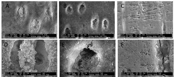
Figure 2.

Electron micrographs of the dentin of the deciduous teeth. The imagens were obtained by scanning electron microscopy at 5,000x (A, B, C) and 20,000x (D, E, F) magnifications. A, D—nonirradiated dentin; B, E—irradiated dentin (30 Gy); C, F—irradiated dentin (60 Gy).
