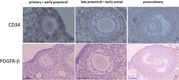
Figure 2.

Immunohistochemical detection of CD34 (a vascular endothelial cell marker) and PDGFR-β (a pericyte marker) in the parabiosis model. Six to twelve ovaries were obtained from three to six wild-type mice in their natural estrous cycles. Immunostaining was evaluated on three to four tissue sections in each ovary in each developmental stage of the follicles. The developmental stages are defined in Materials and Methods. Scale bars; 50 μm.
