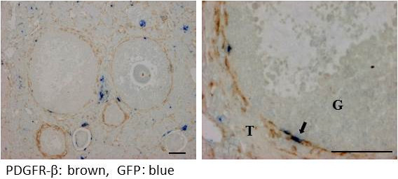
Figure 6.

Double immunostaining for PDGFR-β (a pericyte marker) and GFP (a bone marrow derived-cell marker) in the parabiosis model. Brown shows PDGFR-β and blue shows GFP. Double-positive cells are indicated by arrows. T: theca cell layer, G: granulosa cell layer. Scale bars; 50 μm.
