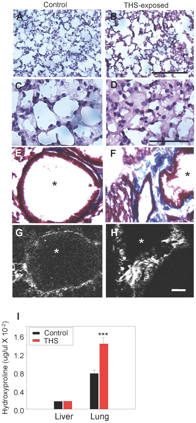
Figure 3. THS exposure results in excess deposition of collagen in lungs.
Cross-sections through the lungs show that in THS-exposed animals, the alveoli in the region of the alveolar sacs are disrupted in comparison to the control animals (A,B). In the terminal respiratory bronchioles of the lung, however, the walls of the alveoli in the THS-exposed animals are thicker and appear to contain secretions (C,D). (E–F) Masson-trichrome staining for fibrillar collagen (blue) shows that the level of collagen in normal lung is low but THS-exposed animals show higher levels of fibrillar collagen with disrupted structure between alveoli (*). (G,H) Second-harmonic imaging microscopy (SHIM) confirms that collagen between alveoli (bright white) remains fibrillar in THS-exposed animals. (I) Hydroxyproline (an amino acid that is highly present in fibrillar collagen) is much higher in lung tissue of THS-exposed animals than in the control. Alveoli in E–H marked by *. Scale bar in A,B is 100 µm, in C,D is 50 µm, in E–H is 20 µm. In A–H and in I. *** p<0.001.

