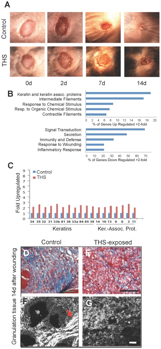
Figure 5. THS exposure delays closure of and weakens cutaneous wounds.
(A) Representative excision wounds performed on the backs of mice after hair removal show that THS exposure results in keratinization of the epithelium (crusty appearance) and delayed wound closure. (B) At day 14, when control wounds are closed, gene expression evaluated by Affirmetrix Arrays shows that THS exposure upregulates keratin genes and genes involved in epithelial migration and contractile function of wound tissue and downregulates genes involved in inflammatory and immune responses. (C) The upregulated keratin genes are primarily associated with hair and nail production, resulting in stiffening of the wound, making it crusty and potentially more fragile. “Ker.-Assoc. Prot.” = Keratin-Associated Protein. (D–G) In cross sections through the healing tissue, Masson-trichrome staining shows great decrease of interstitial collagen (blue) in THS-exposed tissue; second-harmonic imaging microscopy (SHIM) shows that the collagen is strongly fibrillar in the control (arrow) but not at all fibrillar in THS-exposed mice. For A; for B,C, for D–G. Scale bar for D,E = 100 µm and for F,G = 20 µm. White * labels a hair follicle.

