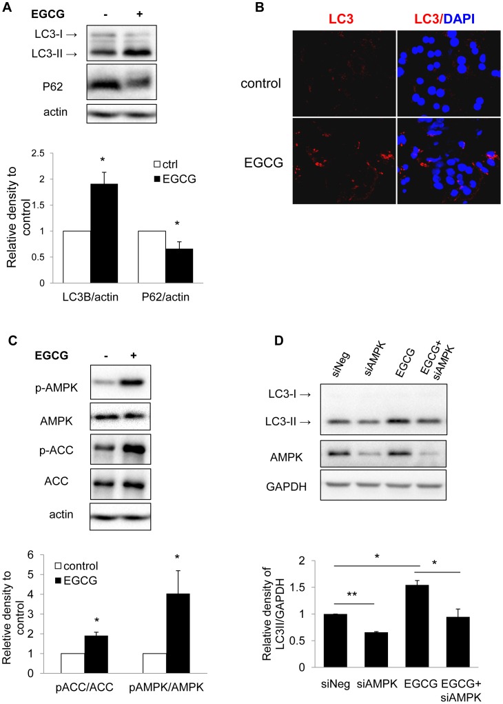
Figure 3. EGCG increase autophagy flux and AMPK phosphorylation in primary hepatocytes.
(A) Immunoblots and densitometric analysis showing changes in LC3-II and P62 level in mouse primary hepatocytes cells treated with 40 µM EGCG for 24 h. Bars represent the mean of the respective individual ratios±SD (n = 3). (B) LC3 immunostaining showing increased endogenous LC3-II puncta in primary mouse hepatocytes treated with 40 µM EGCG for 24 hrs. (C) Immunoblots and densitometric analysis showing changes in p-AMPK, and p-ACC level in mouse primary hepatocytes cells treated with 40 µM EGCG for 24 h. Bars represent the mean of the respective individual ratios±SD (n = 3). (D) HepG2 cells were transfected with negative or AMPK siRNA and incubated for 24 hr. The cells were then treated without or with EGCG (40 µM) for another 24 hr. Bars represent the mean of the respective individual ratios±SEM (n = 3).

