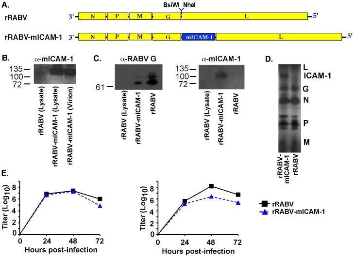
Figure 1. Construction, recovery and characterization of a recombinant RABV-based vaccine expressing the murine Icam1 gene (rRABV-mICAM-1).
A) At the top is a parental recombinant RABV-based vaccine containing an additional transcription stop-start signal flanked by two unique restriction sites between the G and the L genes. rRABV, which is a molecular clone of the SAD-B19 vaccine strain of RABV, was the target to introduce the gene encoding murine ICAM-1. B). BSR cells, which are not expected to express ICAM-1 or its binding partner LFA-1, were infected with rRABV or rRABV-mICAM-1 and lysed 48 hours later. Proteins were separated by SDS-PAGE and subjected to Western blotting with antibodies specific for ICAM-1. A protein of the expected size for ICAM-1 was detected from lysates of rRABV-mICAM-1-infected but not from rRABV-infected BSR cells. In parallel, sucrose-purified and concentrated virus was analyzed by western blot analysis and a protein of the expected size for ICAM-1 was detected in the virion particle. C) Sucrose-purified rRABV-mICAM-1 was re-purified using 5% sucrose step gradient and the single band was collected and analyzed by Western blot analysis to show ICAM-1 protein migrated within the sucrose gradient at the same density as rRABV. RABV and lysates from uninfected BSR cells served as controls for the Western blot analyses. D) Silver staining of separated proteins from sucrose step gradient-purified particles shows a protein at the expected size of ICAM-1 and that the incorporation of ICAM-1 into the virus particles does not affect viral protein compositions. E) BSR cells were infected with rRABV or rRABV-mICAM-1 at a MOI of 5 (one step growth; left) or .01 (multi-cycle growth curve; right). Aliquots of tissue culture supernatants were collected, and viral titers were determined in duplicate.

