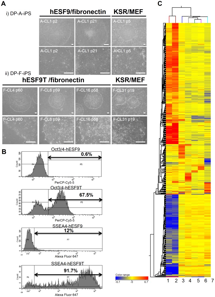
Figure 4. hiPSCs derived from DPCs in completely serum- and feeder-free culture conditions.
A) Phase contrast images of iPSCs derived from DPCs (DP-A-iPS and DP-F-iPS). i) DP-A-iPS-CL1 at passage 2, or 21 on fibronectin-coated dish with hESF9 medium. Right panel showed the cells at passage 5 cultured on MEF with KSR-based conditions. ii) DP-F-iPS-CL4 at passage 60, CL6 at passage 59 and CL16 at passage 58 on fibronectin-coated dish with hESF9T medium. Right panel showed CL31 at passage 19 on MEF with KSR-based conditions. Bars indicate 200 µm. B) Flow cytometry analysis of Oct3/4 and SSEA-4 expression in hiPSCs generated and maintained in hESF9 medium supplemented with TGF-β1 (2 ng/ml) (hESF9T) or without TGF-β1 (hESF9) (DP-F-iPS-CL-8 at passage 33). The horizontal bar indicates the gating used to score the percentage of cells antigen positive. C) Comparison of the global gene expression analysis. Unsupervised clustering was performed using microarray data from parental cell (DPCs), DP-iPSCs cultured in hESF9 or hESF9T (DP-A-iPS, DP-F-iPS) and hiPSCs (Tic, DP-F-iPS). 1.DP cell (DP-A): passage 2 = before infection. 2.DP cell (DP-F): passage 4 = before infection. 3.DP-A-iPS-CL1: passage 14 = serum-free condition (hESF9/on FN). 4.DP-F-iPS-CL12: passage 36 = KSR-based condition (KSR/on MEF). 5.DP-F-iPS-CL6: passage 37 = serum-free condition (hESF9T/on FN). 6.DP-F-iPS-CL8: passage 35 = serum-free condition (hESF9T/on FN). 7.Tic (hiPSC: JCRB1331): passage 58 = KSR-based condition (KSR/on MEF). A genome-wide gene expression profiling analysis demonstrated that hiPSCs cultured in hESF9 or hESF9T on fibronectin showed a similar gene expression pattern to those grown in a conventional feeder-dependent culture (KSR-based condition). Hierarchical combined tree on compare. (Fold change> = 20).

