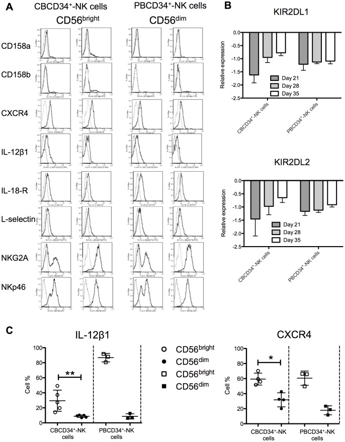
Figure 4. Phenotype of NK cells differentiated from CBCD34+ and PBCD34+ cultures.
(A) Surface antigens were detected by flow cytometry. A representative sample for the expression of each antigen in the CD56bright and CD56dim subsets for CBCD34+ and PBCD34+ cultures is shown. (B) Transcriptional analysis for KIR2DL1 and KIR2DL2 at days 21, 28 and 35 is shown for NK cells from CBCD34+ (n = 4) and PBCD34+ (n = 3) cultures. Values are normalized using three reference genes. (C) Graphs depict the surface expression at day 35 of IL-12β1 and CXCR4 on NK cell subsets from frozen CBCD34+ (n = 4–5) and PBCD34+ (n = 3) cultures. Mann-Whitney test was performed, * P<0.05, ** P<0.005.

