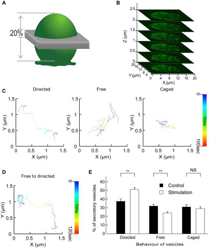
Figure 1. Acquisition and tracking of secretory vesicles in chromaffin cells and categorization of their motion.
Chromaffin cells expressing hGH-GFP were imaged by confocal microscopy. (A) Six optical slices were acquired to obtain a 3 µm z-stack encompassing 20% of the total height of a chromaffin cell (B). (C) Time-coded prototypical trajectories of vesicles tracked for 100 sec illustrating the three different types of vesicular movement as indicated. (D) Time-coded prototypical vesicle trajectory showing a switch from caged behaviour (blue) to directed motion. (E) Comparison of percentages of vesicles in each of the three different motion pools in control conditions and during nicotine (10 µM) stimulation (N = 7 cells, n = 1159 tracked vesicles). Note the significant increase in directed motion and the parallel decrease in the percentage of vesicles undergoing free diffusion. **p<0.01 (paired t-test), NS: not significant.

