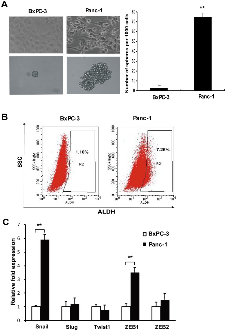
Figure 1. Differences of epithelial-mesenchymal features and CSC properties in Panc-1 and BxPC-3 cells.
A. Morphology of Panc-1 and BxPC-3 cells and their spheres. Note that Panc-1 cells have more spindle-shaped mesenchymal populations and can form more and larger spheres. ** P<0.01 compared with BxPC-3. B. ALDH activity in Panc-1 and BxPC-3 cells. Dot plots of cells analyzed by flow cytometry for ALDH activity. Cells were treated with Aldefluor substrate in the presence or absence of ALDH inhibitor DEAB. After treatment, the samples were analyzed by flow cytometry for the presence of ALDHhigh cells. The values presented are the averages of three independent experiments. C. Real-time RT-PCR quantifing Snail, Slug, Twist1, ZEB1, and ZEB2 mRNA expression in Panc-1 and BxPC-3 cells. Bar graphs show the ratio of the expression level in Panc-1 cells to that in BxPC-3 cells. ** P<0.01.

