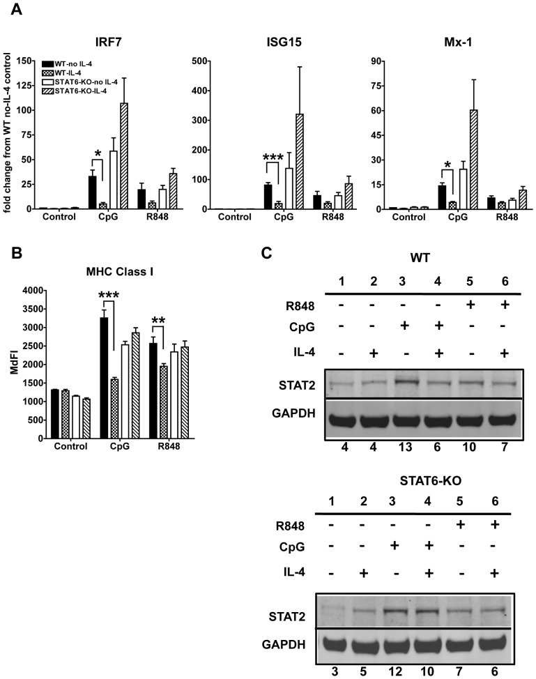
Figure 5. IL-4 suppression of TLR7- and TLR9-induced IFN-dependent response in cDCs is STAT6 dependent.
A. We analyzed by qPCR the gene expression of the IFN-responsive genes IRF7, ISG15 and Mx-1 induced by 6 h stimulation with CpG or R848 in wild type (WT) (C57BL/6) and STAT6-KO cDCs treated or not for 24 h with IL-4. Results were normalized to WT control cells not treated with IL-4. B. We analyzed the MHC Class I expression on cDCs by flow cytometry. Results are shown as median fluorescence intensity (MdFI). Averages and SE of three independent BMDC cultures are shown. (* p<0.05,**p<0.01, ***p<0.0001). C. Western Blot analyses of STAT2 expression in cDCs treated or not for 24 h with IL-4 and harvested 8 h after stimulation with CpG or R848 in WT and STAT6-KO BMDCs. GAPDH was used as loading control. One blot representative of three independent experiments is shown. Numbers below the blot represent the percentage of the normalized integrated density values against GAPDH (loading control).

