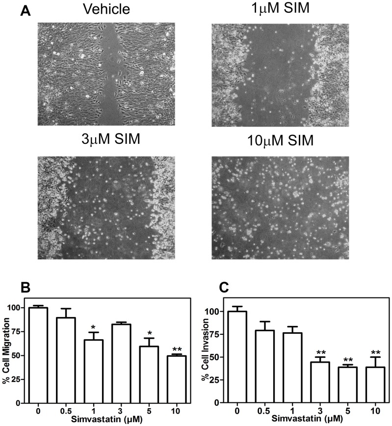
Figure 3. Migration and invasion levels of Simvastatin UMR-106 treated cells.
UMR-106 monolayer were scraped with a pipette tip and incubated in the absence or presence of simvastatin. Wounds were photographed at 48 h after treatment and a representative result of three experiments is shown to schematize the inhibitory effect of simvastatin on migration rate (A). Cells were seeded in the upper level of 8 µm-Boyden chamber uncoated (B) or coated with matrigel (C), attracted by simvastatin and 10% FBS supplemented medium in the lower chamber. After 48 h migrating cells were stained with crystal violet methanol solution, the 8 µm-pore membranes were cut off and crystals were dissolved in 10% acetic acid. Absorbance solution was measured at 540 nm. Quantitative response was expressed as percentage of the control ± SEM of two replicates. a means p<0.001, b means p<0.05 vs control, after a one-way ANOVA analysis.

