
Figure 2. Distribution of dextran-labeled vagal fibers in the mesenteric ganglion and spleen coronal section.
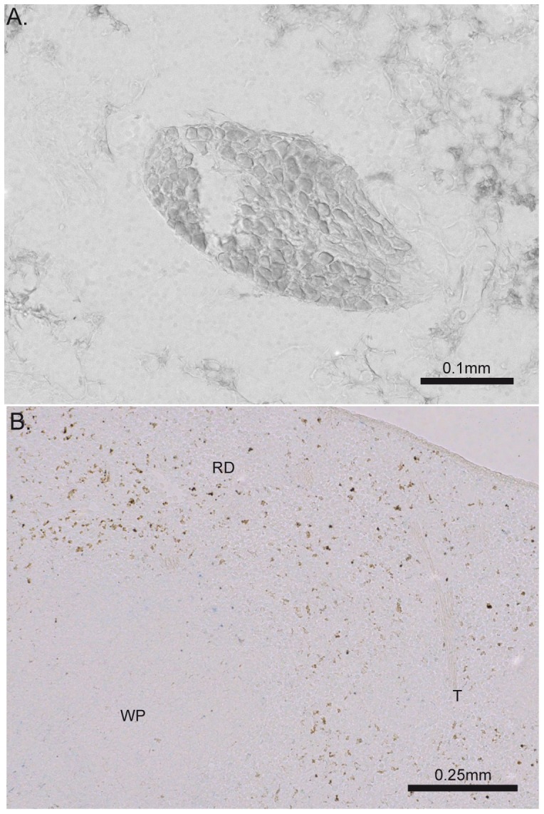
No biotin dextran-labeled fibers or terminals were found on coronal section of mesenteric ganglion (A) or spleen (B). Of note, similar observations were obtained with Texas red dextran amine tracer. The brown spots on the spleen section were found in injected and non-injected mice, indicating of a strong endogenous biotin expression. WP: white pulp, RP: red pulp, T: trabeculae.