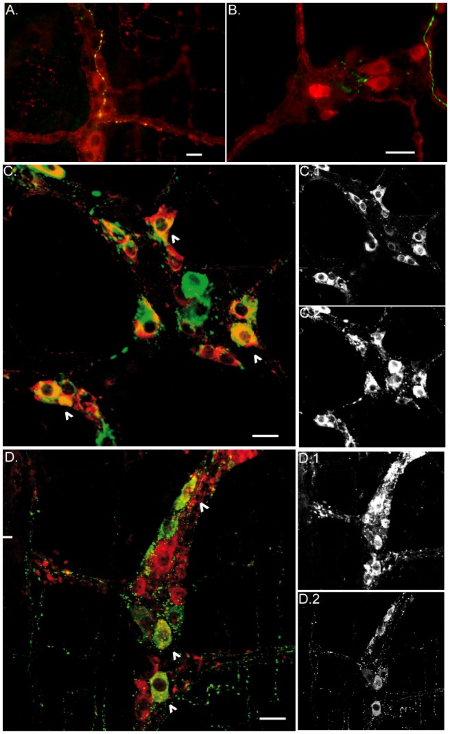
Figure 7. Vagus nerve efferent fibers and terminals are close to cholinergic and nitrergic enteric neurons.
A. Epifluorescence image shows dextran-labeled vagal efferents (green) that co-localize with ChaT (yellow), and are in close contact with ChaT positive enteric neurons (red). B. Epifluorescence image shows labeled vagal efferent fibers (green) making contact with nNOS positive neurons (red). Of note, cholinergic neurons, and to lesser extent nitrergic neurons, are the main population targeted by the vagal efferent fibers. C. Confocal image of VIP (red) and nNOS (green) myenteric neurons. Most of the cells bodies exhibit co-localization of these two neurotransmitters (arrow head). D. Confocal image of VIP (red) and ChaT (green) myenteric neurons. Occasionally myenteric neurons showed immunoreactivity for both neurotransmitters (arrow head). C1 and C2 show the distribution for the nNOS and VIP positive cells bodies, respectively. D1 and D2 show the distribution for ChaT and VIP positive cells bodies. Scale bar represents 20 µm.

