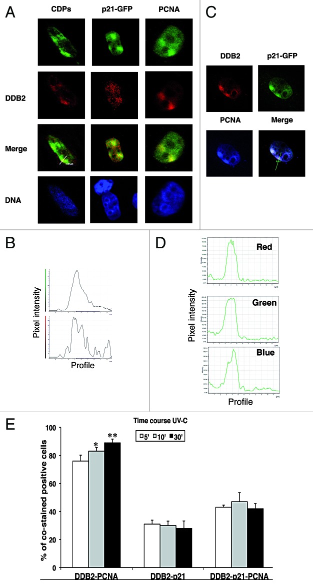
Figure 1. Co-localization of DDB2-NER proteins at DNA damaged sites. HeLa cells were co-transfected with DDB2 and p21wt-GFP constructs. For color images, refer to the online version of the article. (A) Twenty-four h after transfection, cells were exposed to local UV-C irradiation (100 J/m2) through filters with 3-μm pores, extracted in situ and then fixed for immunofluorescence staining with anti-DDB2 (red fluorescence) and anti-CDPs or anti-PCNA antibody (green fluorescence; p21 protein is detectable thanks to fusion with GFP). DNA was counterstained with Hoechst 33258 (blue fluorescence). Representative confocal merged images show the co-localization with the DDB2 protein. (B) The distribution profiles of the pixel intensity of the green and red channels along the white line drawn in the merged image show the co-localization of DDB2 with CDPs at the DNA damaged sites. (C) Confocal microscopy sections of irradiated HeLa cells showing DDB2 (red signal), PCNA (blue signal), p21 (green signal). The merged image proves the co-localization of the 3 proteins. (D) The distribution profiles of the pixel intensity for each fluorescence signal (blue, green, and red) analyzed by confocal microscopy are shown. (E) HeLa cells co-transfected with p21wt-GFP and DDB2 expression vectors and 24 h later exposed to UV-C radiation (100 J/m2). The cells were extracted in situ and fixed for determining the percentage of cells showing co-localization of DDB2-PCNA, DDB2-p21, and DDB2-PCNA-p21. Cells were analyzed at 5 min (empty bars), 10 min (gray bars), and 30 min (black bars) after UV-C irradiation.

An official website of the United States government
Here's how you know
Official websites use .gov
A
.gov website belongs to an official
government organization in the United States.
Secure .gov websites use HTTPS
A lock (
) or https:// means you've safely
connected to the .gov website. Share sensitive
information only on official, secure websites.
