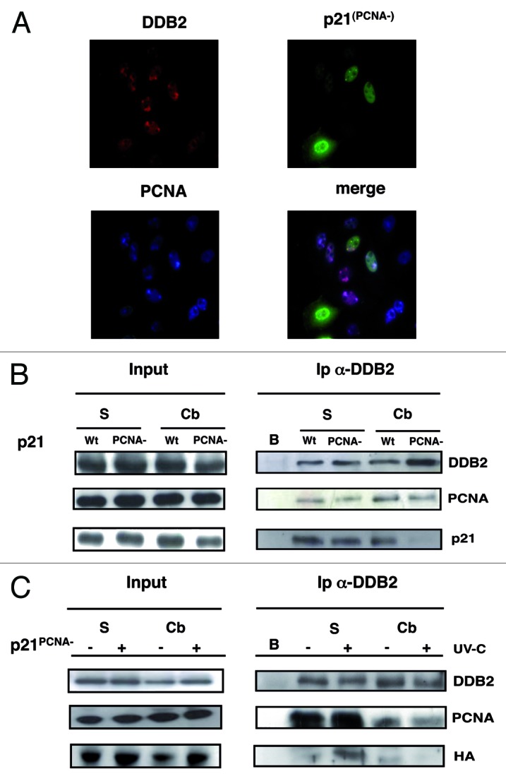
Figure 3. Interaction of DDB2 with p21 is mediated by PCNA. For color images, refer to the online version of the article. (A) HeLa cells grown on coverslips were lysed in situ and fixed 30 min after UV irradiation, as described in “Materials and Methods” for immunofluorescence analysis of chromatin-bound proteins: DDB2 (red fluorescence), PCNA (blue fluorescence), and HA-p21PCNA– (green fluorescence). (B) HeLa cells expressing exogenous DDB2 protein and HA-p21wt or HA-p21PCNA– were collected 30 min after UV-C irradiation (30 J/m2). Immunoprecipitation (Ip) with anti-DDB2 antibody from soluble (S), chromatin-bound (Cb) fractions. Not transfected cells (sample B). Input load: 1/30 of cell extracts. (C) Immunoprecipitation (Ip) with anti-DDB2 antibody of soluble (S), chromatin-bound (Cb) samples obtained before or after 30 min UV-irradiation (30 J/m2) from cells expressing DDB2 exogenous protein and HA-p21PCNA–. Not transfected cells (sample B). Input load: 1/30 of cell extracts.
