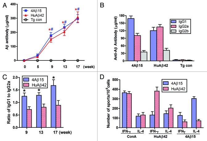
Figure 1. Generation of immune responses in APP/PS1 mice immunized with 4Aβ1-15 peptide plus MF59. (A) Aβ antibody titers were measured by ELISA. Data are presented as mean ± SD of Aβ antibodies (μg/ml). One-way ANOVA followed by post hoc comparison revealed significant differences in anti-Aβ titers of 4Aβ1-15-immunized group when comparing week 9 to weeks 13 or 17 (n = 9, *P <0.01). The same trend was observed in human Aβ42-immunized group (n = 9, #P < 0.01). (B) Detection of IgG1, IgG2a and IgG2b subclasses of anti-Aβ antibodies in mice immunized with the vaccines. Isotyping in sera from immunized mice after the final immunization. (C) The results revealed significant differences of the ratio of IgG1 and IgG2a between 4Aβ1-15 vs. Aβ42-immunized group at each week shown (n = 9, *P <0.01). (D) Lymphocytes from APP/PS1 mice immunized with Aβ42 or 4Aβ1-15 were individually isolated and cultured then stimulated with ConA (5 μg/ml), 4Aβ1-15 or Aβ42 (20 μg/ml) for 36 h. The number of IL-4 and IFN-γ positive T cells in immunized mice detected by ELISPOT. Data were presented as mean ± SD of each cytokine. There was no significant difference between vaccine-treated groups for levels of each cytokines after in vitro ConA challenge. However, there was a significant difference between groups in cytokine levels of IL-4 and IFN-γ after 4Aβ1-15 or Aβ42 challenge.

An official website of the United States government
Here's how you know
Official websites use .gov
A
.gov website belongs to an official
government organization in the United States.
Secure .gov websites use HTTPS
A lock (
) or https:// means you've safely
connected to the .gov website. Share sensitive
information only on official, secure websites.
