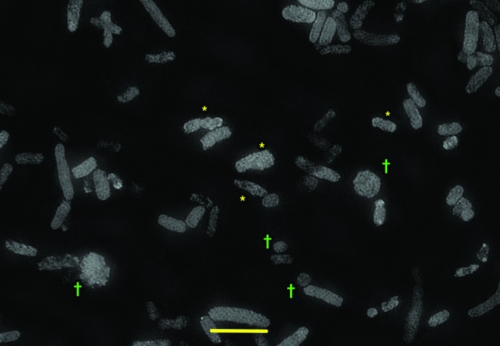
Figure 1. Visualization of the envelope stress response in Escherichia coli strain E2348/69. Classic enteropathogenic E. coli strain E2348/69 was grown in DMEM medium in the presence of 0.1 mg/L trimethoprim in order to increase the size of the E. coli cells to improve visualization. Trimethoprim alone did not cause any changes in the E. coli cell shape or contour (images not shown). After 1 h of growth, zinc acetate was added to 0.4 mM final concentration and growth was continued for 3 more h (4 h total). At this time the E. coli bacterial suspension was mixed with an equal volume of 0.2% acridine orange in ethanol and allowed to stain at room temperature for 10 min. Staining was followed by two cycles of washes consisting of centrifugation at 700 g x 10 min, followed by decanting and resuspension in water. After the second wash, 40 µl of bacterial suspension was spotted on an ordinary glass microscope slide and allowed to dry. The slides were sent to Adrian Quintanilla of the Applied Precision division of GE Healthcare, Issaquah, WA, who performed the imaging using the OMX instrument and the Structured Illumination Microscopy protocol. Color images were converted to black and white. *E. coli cells showing distorted shapes, bumpy cell outlines, and moth-eaten appearance. †E. coli cells showing spheroidal shapes. Size bar at bottom is approximately 4 µm.
