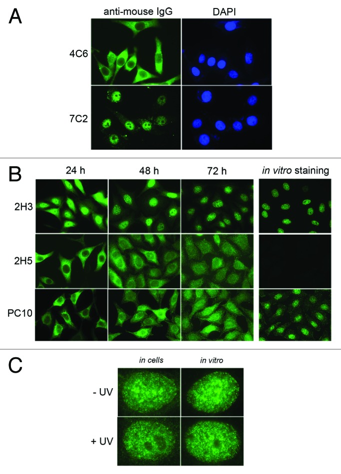
Figure 1. Intracellular localization of delivered antibodies as probed by immunofluorescence microscopy. (A) Micrographs show typical HeLa cells transduced with either 4C6 or 7C2 antibody. The delivered antibodies were revealed with Alexa 448-labeled goat anti-mouse immunoglobulins 24 h post-transduction. Both antibodies are IgG1κ. Magnification: 630 × . (B) The pictures correspond to typical fields of HeLa cells transduced with the indicated antibodies at 24, 48 and 72 h post-treatment. The delivered antibodies were revealed as in A. The panels on the right correspond to the staining by indirect immunofluorescence of HeLa cells incubated with the relevant antibody (in vitro staining). Magnification: 630 × . (C) The pictures show typical nuclei of HeLa cells either transduced and fixed at 48 h post-treatment (in cells) or stained by indirect immunofluorescence (in vitro) with 2H3 antibody without (- UV) or with (+ UV) UVC irradiation 8 h before analysis. Magnification: 100 × .
