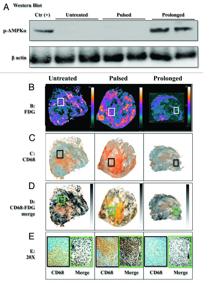
Figure 5. Pictures from harvested tumors. (A) displays western blot data showing that increased phosphorylation of AMPK only occurred in animals exposed to prolonged treatment. (B) display a large macrophage infiltration in pulsed group as detected by double staining immunohistochemistry for CD68 antigen (brown) and cytokeratin CK19 (purple). (C) display autoradiography of FDG uptake, while coregistration of metabolic and immunohistochemical data are reported in (D) and a magnified area (20×) in (E). Co-localization of tracer retention and CD68+ cells can be best appreciated after pulsed treatment, while it is less evident in the remaining specimens.

An official website of the United States government
Here's how you know
Official websites use .gov
A
.gov website belongs to an official
government organization in the United States.
Secure .gov websites use HTTPS
A lock (
) or https:// means you've safely
connected to the .gov website. Share sensitive
information only on official, secure websites.
