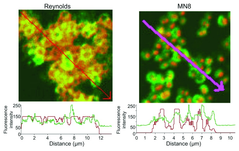
Figure 2. Quantification of the fluorescence intensity of the reactivity of S. aureus CP5 strain Reynolds and CP8 strain MN8 to antibody to the homologous CP antigen and PNAG. Binding of primary rabbit antibody to purified CP5 or CP8 conjugate antigens to S. aureus cells detected with anti-rabbit IgG secondary antibody conjugated to AlexaFluor (AF) 588 (red). Human mAb F598 to PNAG directly conjugated to AF 488 (Green) was used to detect PNAG on the bacterial surface. Histograms depict analysis of the co-localization of red and green pixels in samples reacted with antibody to both CP and PNAG antigens across the distances, in microns (µm), depicted on the X-axis. Arrows on photomicrographs indicate regions analyzed.

An official website of the United States government
Here's how you know
Official websites use .gov
A
.gov website belongs to an official
government organization in the United States.
Secure .gov websites use HTTPS
A lock (
) or https:// means you've safely
connected to the .gov website. Share sensitive
information only on official, secure websites.
