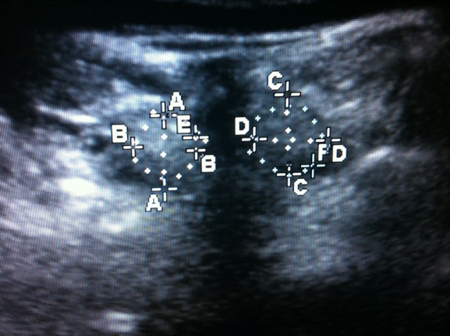
Fig. 1.

Ultrasound image in the transverse plane with multifidus muscle encircled either side of the posterior spinous process. Anteroposterior (AA, CC) and mediolateral (BB, DD) dimensions and cross-sectional area (CSA: E, F) from both left and right sides were added together to represent CSA at each level
