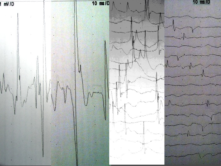
Fig. 3.
a–d Electromyography examples in study patients. a Normal. b Mild chronic change, high amplitude wide complex unit adjacent to normal unit. c Moderate subacute neurogenic change with ongoing prominent fibrillations, moderately reduced interference pattern suggesting ongoing denervation with reinnervation. d Severe neurogenic abnormality with widespread fibrillation potentials

