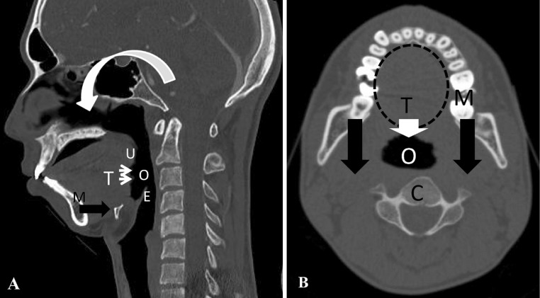
Fig. 1.
Schematic drawing of the proposed mechanism of oropharyngeal stenosis. U uvula, E epiglottis, M mandible, T tongue root, O oropharynx, C cervical spine. a Sagittal computed tomography reconstruction of the cervical spine. Reduction in the O-C2A flexes the maxilla (curved white arrow), which makes the mandible shift posteriorly (black arrow) with the tongue root (thin white arrows). b Movement of the tongue root (while arrow) caused by the backward shift of the mandible (black arrow) results in oropharyngeal stenosis

