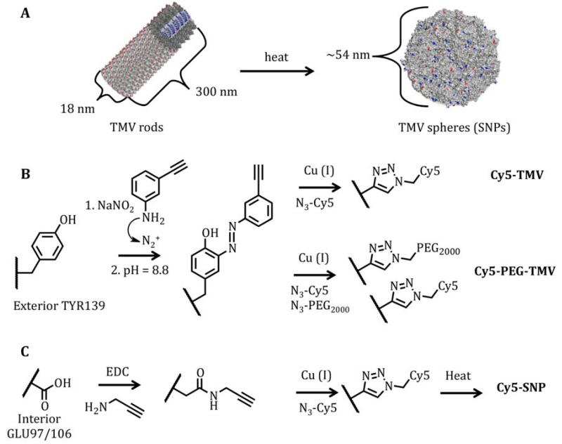
Figure 1.
(A) A PyMol generated schematic representation of the thermal transition of tobacco mosaic virus (TMV) rods to spherical nanoparticles (SNPs). Several coat proteins have been removed from the TMV rod to highlight the hollow interior with interior glutamic acids, GLU97 and GLU106, in blue and exterior tyrosine, TYR139, in red. (B) Schematic illustration of the bioconjugation reactions used to incorporate sulfo-Cy5 dyes and PEG2000 molecules on the exterior of TMV rods. (C) Schematic illustration showing the method to generate sulfo-Cy5 labeled SNPs.

